Booking Information
The department houses both a transmission (TEM) and scanning electron microscope (SEM). Using electrons with much smaller wavelengths than visible light, electron microscopes are capable of resolving smaller features than light microscopes, in the nanometer to angstrom ranges depending on the configuration of the electron microscope.
SEM can accommodate most materials for analysis. The TEM sample holders are designed to accept 3 mm diameter disks. Samples need to be electron transparent (usually < 100 nm thick).


To request instrument time for the microscopy facility, please visit our online booking software, QReserve, and follow these steps:
- After selecting the instrument you wish to request, you will be asked to log in with your Queen’s ID.
- Select the Reserve Now button near the top of the page and fill out the form to submit your request.
- Students/Researchers can be trained to run their samples independently. The request is also made through QReserve. Upon successful completion of the training program the user will have access to the instrument calendars for booking future appointments.
Not affiliated with Queen's? Contact microscopy@queensu.ca.
SEM:
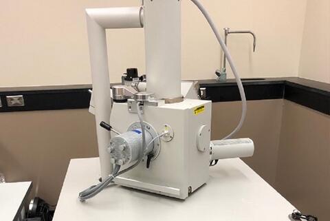
Thermo Fisher Quanta 250 eSEM with Everhart-Thornley, Concentric Backscattered Electron, Large Field, and Gaseous Secondary Electron detectors for variable pressure measurement of both secondary and backscattered electrons. An EDAX Element EDS detector is used for elemental characterization including mapping.
TEM:
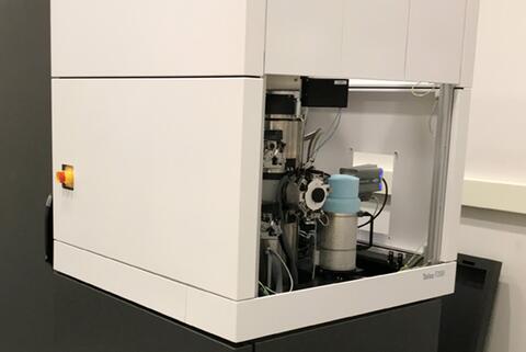
Thermo Fisher Talos F200i S/TEM with single-tilt, double-tilt, tomography, and cryo-tomography holders. X-TWIN polepiece for high angle tilt capabilities with a constant power objective lens. 16 Mpixel camera for imaging/diffraction work capable of 40 fps for dynamic experiments. HAADF and on-axis BF/DF2/4 solid state detectors for annular collection of scattered electrons in scanning mode. Bruker Quantax EDS detector allows elemental identification and mapping.
SEM can accommodate most materials for analysis.
- Samples should be vacuum compatible (1 torr and below, typically 10-4 torr) and free of loose particles.
- Conductive samples are easier to image but non-conductive samples can be run either by coating with a thin conductive layer or using one of the low vacuum modes to minimize sample charging.
- Samples should be able to fit on a ¼” diameter stub, the stage can hold 7 stubs for multiple sample imaging.
- Samples should be dry, preferably placed under vacuum prior to entering the SEM chamber.
- Powder-type samples can be pressed into a pellet or deposited on double-sided conductive tape. Any loose particles should be removed using clean air prior to introduction into the vacuum chamber.
The TEM sample holders are designed to accept 3 mm diameter disks. Samples need to be electron transparent (usually < 100 nm thick) to be
- Samples need to be vacuum compatible (10-7 torr and lower).
- Samples generally do not need to be conductive but coating with a fine layer of metal can help in some applications.
- Particle samples/cut samples can be supported on metal grids with or without a film coating on the grid.
- Sample preparation, Sample preparation, Sample preparation. Sample preparation is key in determining the success of TEM. The department is building its suite of sample preparation equipment and the Facility Manager can assist in determining the correct tools for your sample preparation.
Samples will be disposed of 30 days after completion of the analyses, unless the return of the samples is requested on the Request Form. Sample return costs will be added to your invoice unless a courier account number (FedEx or DHL) is provided.
Scanning Electron Microscope (SEM)
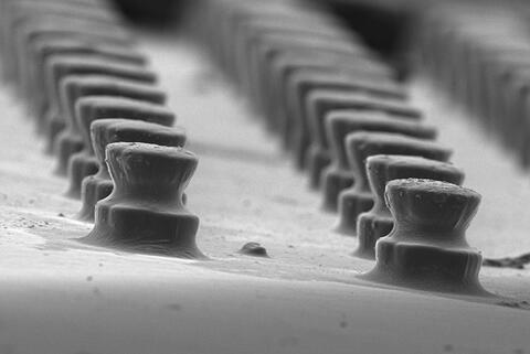
In SEM a beam of electrons (~1-30 kV in energy) is scanned across the surface of the sample and information collected at each point (pixel) of the sample. This gives topographical and elemental information (when coupled with energy dispersive X-ray spectroscopy) from the sample at relatively low to moderate magnifications (25x - ~100,000x).
Our Quanta 250 environmental SEM is equipped with detectors for both secondary and backscattered electrons at a range of vacuum pressures, with an EDAX Element EDS detector for elemental X-ray characterization of samples. Samples should be conductive or can be coated with a conductive layer. However, the use of a variable pressure chamber allows for the imaging of non-conductive samples as well.
Transmission Electron Microscope (TEM)
By contrast, a TEM functions similarly to a light microscope where the whole area of interest is illuminated by the beam and the image is magnified by lenses to be projected on a camera. TEM utilizes higher energy electrons (~40-200 kV in energy) than SEM which allows much higher magnifications to be achieved (up to ~ 1,000,000x) but also requires very thin samples (for electron transparency). Scanning coils incorporated in the lens column of the TEM allows even greater magnifications (~10,000,000x) in scanning transmission electron microscopy (STEM) mode for very high-resolution work. The addition of an energy dispersive X-ray detector (EDS) affords elemental characterization in both the SEM and STEM.
Our Talos F200i scanning TEM has multiple sample holders, single and double tilt for routine imaging and diffraction, a high visibility tomography holder for 3D volume reconstructions, and a high tilt cryo-holder for biological and sensitive samples. The Talos is equipped with a 16-megapixel camera with a capability of 40 frames per second for dynamic experiments. In STEM mode various angular distributions of scattered electrons are collected on the on-axis brightfield/segmented dark field detectors and high angle annular dark field detector. A Bruker Quantax energy dispersive spectrometer is used for elemental characterization of samples in scanning mode. Samples need to be thinned to thicknesses around 100 nm before imaging in the TEM.
| Address
Department of Chemistry |
Fax
613-533-6669 |
Hours
Monday to Friday |
Contact Information
Email: microscopy@queensu.ca